Your growth scan is one of the common ultrasound examinations you are likely to undergo during your pregnancy, between 20 – 40 weeks. The main objective is to check on how well your baby is growing. But if you haven’t had this scan before, you may not know what to expect or what the purpose of the scan is. Here’s some information to help you be prepared.
Medical reasons for this scan include the following:
- Previous history of small baby
- Bleeding in pregnancy or abdominal pain
- Pregnancy with twins
- Medical problems e.g. gestational diabetes, high blood pressure etc.
- Reduced baby movements
- Suspicion of baby measuring too small or too big
- Abnormal presentations e.g. breech
- Abnormalities of placenta e.g. location etc.
- Variety of reasons as observed by doctors and midwives.
What should you bring or do?
There are a couple of things that you need to do when coming for this examination. Firstly, bring your referral which will state the reason for the scan. Secondly, if you have any previous ultrasound reports for the current pregnancy bring these with you for comparison, as they will greatly help in the finalising of your report. Lastly, upon arrival, fill in the questionnaire very carefully at reception. Remember to fill in the expected date of confinement as this will assist to date your pregnancy.
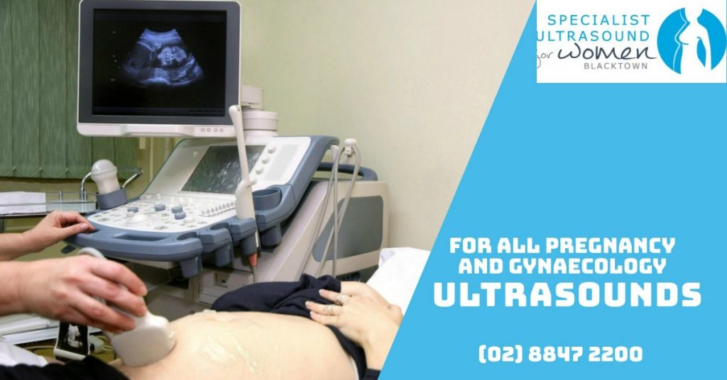
How is this scan done?
This is an abdominal scan most of the time but can be combined with a transvaginal scan to check on placenta and cervix if requested by your doctor or midwife. A moderately full but comfortable bladder is recommended but depending on age of pregnancy may not be necessary. The scan is performed while you are lying on your back. There is a possibility you may feel faint and sweaty during the scan. If this happens notify the sonographer immediately and this can be promptly managed.

What is examined during this scan?
- The baby’s heart rate and rhythm
- Size of baby’s head, tummy and length of the thigh bone. These will help to estimate the baby’s weight.
- Estimation of amount of fluid around the baby and other behavioural baby patterns e.g. breathing, movements etc.
- Blood flow patterns in the umbilical cord and in baby’s brain are checked using Doppler techniques. This gives an indication of wellbeing of placenta.
- Examination of location of placenta and part of baby that is likely to present at time of birth.
- Estimated length of cervix.
- Check for any structural anomaly that may have evolved over time e.g. in the brain, heart, baby’s tummy, kidneys etc.
- 3D and 4D pictures can be taken at the end of the scan.
Your ultrasound report
This will summarise the results of the ultrasound examination. Good baby growth occurs when the placenta is functioning well, leading to provision of adequate oxygen and other nutrients to the baby. The final report will show the following:
- Baby body parts measurements compared to standardised charts.
- Estimated baby weight which will be compared to baby growth charts that can help to detect any growth anomaly patterns.
- Baby wellbeing parameters in umbilical cord and brain blood flow patterns.
- Estimation of fluid around the baby and comments on baby movement patterns.
- Location of placenta and other placental related concerns.
In summary, a growth scan is a very important ultrasound examination in pregnancy if requested by your doctor or midwife. It helps your healthcare provider in guiding them on how to manage your pregnancy. Remember the range of normal baby growth is wide, several scans over time may be needed to determine the trend of growth and ultimately guide management.